Home
 Tell us about your project »
Central New York’s Foremost Faculty, Students and Staff are Available to Help Your Business Innovate.
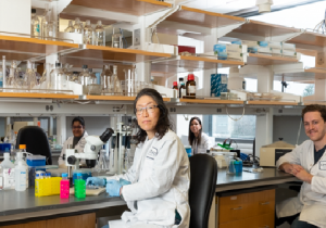
With the resources of the SUNY Research Foundation, and our history of successful partnerships, we are here to help move biomedical products and ideas to market.
Our scientists and core facilities can help move discoveries into practice and technologies into the marketplace.
Upstate is home to top research facilities with highly specialized equipment and advanced instrumentation, to support research and product development.
We are here to create the relationships and partnerships needed to move innovative ideas forward.
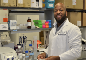
Upstate Biotech Ventures
In a partnership between Empire State Development, Upstate Medical University, the SUNY Research Foundation, and Excell Partners, the newly-launched Upstate Biotech Ventures invests in high-potential startups and small businesses affiliated with Upstate Medical University to drive research and technology innovation.

Recent Tech from SUNY Upstate

This technology employs an engineered self-delivering siRNA to selectively reduce USP10 protein leve...
This technology employs an engineered self-delivering siRNA to selectively reduce USP10 protein levels in tissues, thereby promoting regenerative healing and suppressing pathological scarring. Preclinical studies in the eye demonstrate a favorable safety profile and robust efficacy, supporting the potential of this approach for treating fibrotic disease across multiple organ systems. Background:
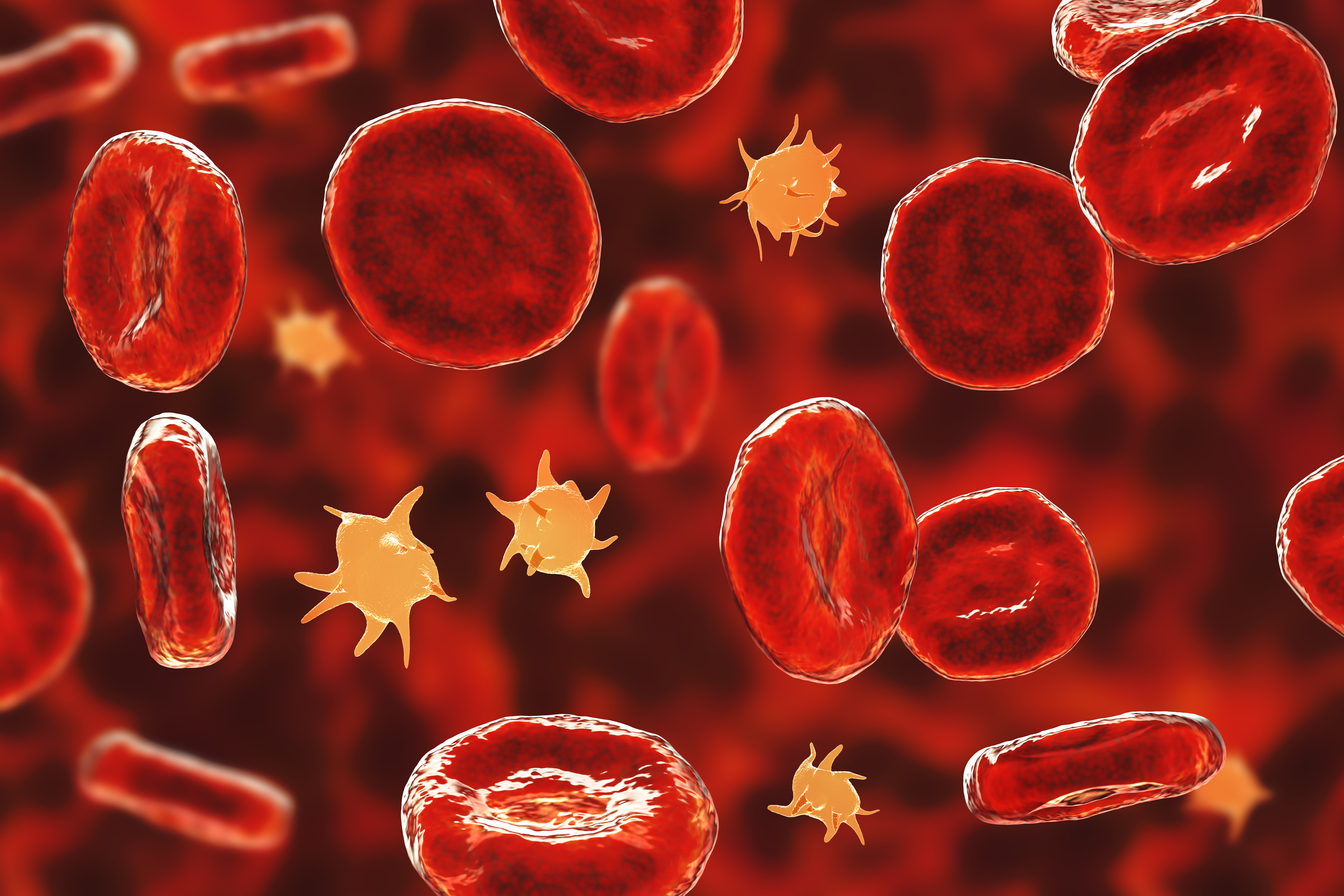
Regenerative healing, particularly in ocular tissues such as the transparent cornea, is a critical area of biomedical research due to the eye’s limited capacity for self-repair and the high risk of vision loss following injury or surgery. Scarring and fibrosis in the cornea can result in permanent visual impairment or blindness, making effective wound healing without scarring a major unmet medical need. Current clinical approaches to treat corneal scarring are limited, with corneal transplantation being the only definitive solution for severe cases. Additionally, in glaucoma surgeries like trabeculectomy, excessive scarring of the surgical site often leads to surgical failure and poor patient outcomes. The need for therapies that can promote regenerative healing while minimizing fibrosis is therefore urgent, not only for ocular health but also for broader applications in tissue repair and organ fibrosis. Existing methods to prevent or treat scarring in the eye, such as the use of antiproliferative agents like mitomycin C, are associated with significant drawbacks, including toxicity, risk of infection, and non-specific inhibition of cell proliferation that can impair normal healing. These treatments do not specifically target the molecular drivers of fibrosis and often result in incomplete or unsatisfactory outcomes. Furthermore, the lack of targeted, non-toxic therapies means that patients frequently require repeated interventions or long-term use of adjunctive medications such as steroids and antibiotics, which carry their own risks and side effects. The limitations of current approaches highlight the need for more precise, effective, and safer therapies that can modulate specific molecular pathways involved in scarring and promote tissue regeneration.Technology Overview:
This technology comprises a self-delivering small interfering RNA (siRNA) therapeutic designed to selectively suppress ubiquitin-specific peptidase 10 (USP10), thereby shifting wound repair toward regenerative healing and away from fibrosis, with particular relevance to corneal and other ocular tissues. The siRNA duplex is chemically modified for enhanced stability, specificity, and delivery efficiency. The technology has demonstrated potent knockdown of USP10 in cell-based assays, significant acceleration of wound closure, and reduction of fibrosis markers in animal models, as well as enhanced epithelial regeneration in ex vivo human corneas. Safety studies in mice, rabbits, and mini-pigs confirmed the absence of toxicity or adverse ocular effects, supporting its suitability for clinical applications. What differentiates this solution is its combination of a novel therapeutic target (USP10) with a highly optimized, self-delivering siRNA platform that achieves effective gene silencing at lower doses and with minimal dosing frequency. Unlike traditional anti-scarring treatments, which often rely on cytotoxic agents like mitomycin C and carry significant side effects, this approach directly modulates the molecular pathways involved in fibrosis and healing, promoting nerve growth, enabling regenerative repair without the risk of immune or vascular complications. The extensive chemical modifications confer nuclease resistance and improved pharmacokinetics, allowing for topical or parenteral administration without the need for complex delivery vehicles. The platform’s efficacy across multiple preclinical models, broad applicability to various fibrotic conditions, and favorable safety profile position it as a transformative advance in regenerative medicine, potentially reducing the need for invasive procedures like corneal transplantation and offering a new paradigm for treating scarring and fibrosis in both ocular and non-ocular tissues. https://suny.technologypublisher.com/files/sites/adobestock_17056179091.jpegAdvantages:
• Promotes regenerative healing and significantly reduces scarring, especially in corneal tissues
• Self-delivery siRNA technology enables effective USP10 knockdown at low doses without immune or vascular reactions
• Demonstrated safety and non-toxicity in preclinical animal models with no adverse effects on corneal morphology or function
• Long-lasting efficacy with effects sustained for at least six weeks post-treatment
• Potential to improve outcomes in ocular surgeries by preventing fibrosis and scarring that lead to vision loss
• Versatile delivery options including topical, pulmonary, and parenteral administration with targeted delivery capabilities
• Broad therapeutic applications beyond ocular healing, including treatment of fibrosis in skin and internal organs
• Reduces need for adjunctive antibiotic or steroid treatments due to its targeted and effective mechanism Applications:
• Corneal wound healing therapy
• Anti-scarring treatment post-eye surgery
• Topical therapy for ocular fibrosis
• Dermal wound regenerative healing
• Fibrosis treatment in internal organs Intellectual Property Summary:
Patent application PCT/US2025/018729 filed on 3/6/2025 Stage of Development:
TRL5
Supported by data appropriate to its current TRL stage, including compelling efficacy in several preclinical models, broad fibrotic disease applicability, and a strong safety profile, the platform stands poised to drive a major advance in regenerative medicine, offering a less invasive alternative to procedures such as corneal transplantation and a novel pathway for treating fibrosis across tissue types.Licensing Status:
This technology is available for licensing.
A serotonin release assay that requires no special training or expensive equipment, and produces re...
A serotonin release assay that requires no special training or expensive equipment, and produces results within hours. Background:
Platelet serotonin release assay (SRA) is a widely-used clinical assay for diagnosing heparin-induced thrombocytopenia (HIT), a life-threatening complication of heparin treatment. The current SRA procedure entails sending patient serum to a facility for testing by radioactive serotonin uptake/release or by mass spectrometry. Both assay methods require specialized equipment and trained personnel, with a turnaround time typically between three to seven days. Unfortunately, the mortality of HIT increases significantly with each passing day. As a result, quicker SRA turnaround time could save lives. Ideally, point-of-care facilities would obtain SRA results within hours, using inexpensive equipment and no special training to perform.Technology Overview:
This technology is a luminescent biosensor that changes from green to blue in the presence of a target analyte. The biosensor consists of two molecules. The first is a specially designed green-to-blue color-changing luminescent protein (nLuc-AFF). The second is one or more short (20-50 nucleotide) DNA hairpins or DNA aptamers of novel design. These bind the target DNA sequence, RNA sequence, small molecule, or protein. Upon binding, the AP-1 sequence becomes exposed and activates the biosensor. The biosensor can be readily adapted to recognize different targets by modifying the DNA component, using existing online DNA design tools. The color change is visible and detection/quantification is via cell phone camera. https://suny.technologypublisher.com/files/sites/adobestock_446068375.jpegAdvantages:
• Produces results within hours.
• Requires no special training or expensive equipment.
• Can be easily adapted to recognize a variety of targets.
Applications:
• Improved platelet SRA for diagnosing HIT.
• Rapid detection of virus or other pathogen infection, such as coronavirus or cytomegalovirus.
• Rapid detection of disease biomarkers such as microRNA, metabolites, or aberrant proteins.
Intellectual Property Summary:
Know-how basedStage of Development:
TRL 3 - Experimental proof of concept Licensing Status:
This technology is available for licensing.Licensing Potential:
This technology would be of interest to anyone involved in the development of methods for detection of HIT and other conditions. These include:
• Pharmaceutics companies.
• Hospitals.
• Medical research laboratories.
• Universities.
This technology uses lung-targeting lipid nanoparticles to deliver a combination of anti-inflammator...
This technology uses lung-targeting lipid nanoparticles to deliver a combination of anti-inflammatory and immune-modulating drugs directly to the lungs, offering a more effective and targeted treatment for acute lung injury and acute respiratory distress syndrome. Background:
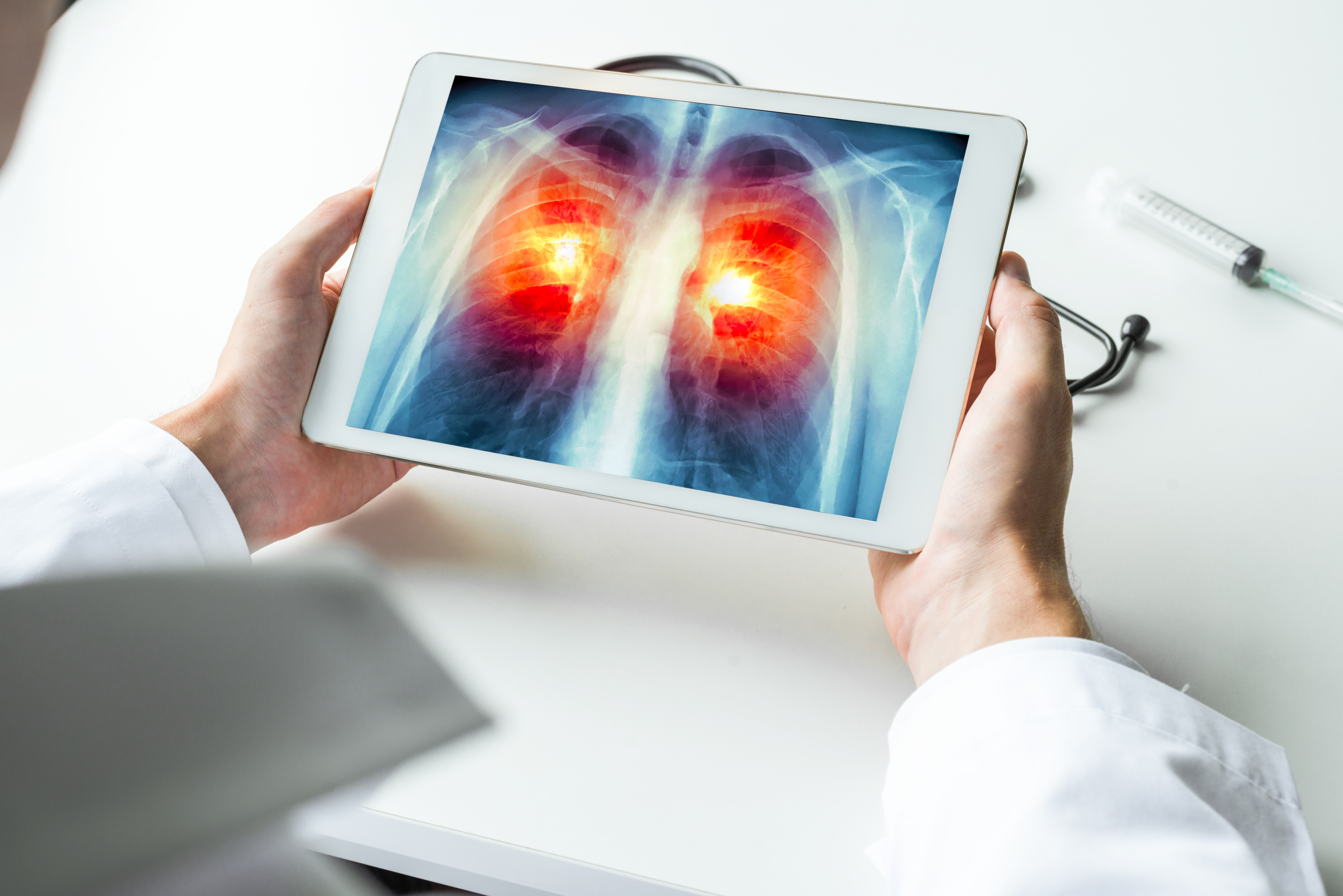
Acute lung injury (ALI) and acute respiratory distress syndrome (ARDS) are severe, life-threatening conditions characterized by widespread inflammation and increased permeability in the lungs, often resulting from infection, trauma, or other critical illnesses. These syndromes lead to impaired gas exchange, hypoxemia, and respiratory failure, frequently requiring intensive care and mechanical ventilation. Despite advances in supportive care, mortality rates for ALI/ARDS remain high, underscoring the urgent need for more effective therapeutic interventions. The complexity of these conditions, which involve dysregulated immune responses and extensive lung tissue damage, has driven ongoing research into targeted therapies that can modulate inflammation and promote tissue repair directly within the lungs. Current treatment strategies for ALI/ARDS are largely supportive, focusing on mechanical ventilation and fluid management, with pharmacological interventions offering only modest benefits. Conventional drugs such as corticosteroids, neuromuscular blockers, and inhaled nitric oxide have shown limited efficacy in improving patient outcomes, and many promising agents—including antioxidants, statins, surfactant therapy, and cytokine inhibitors—have failed to demonstrate consistent clinical benefit. One major limitation of existing approaches is the lack of targeted delivery to lung tissue, resulting in suboptimal drug concentrations at the site of injury and increased risk of systemic side effects. Furthermore, most therapies address only a single aspect of the disease process, rather than the multifaceted immune and inflammatory pathways involved in ALI/ARDS, leaving a significant gap in effective, comprehensive treatment options.Technology Overview:
This technology utilizes specialized lung-targeting lipid nanoparticles (LNPs) designed for the intravenous delivery of multiple therapeutic agents to treat acute lung injury (ALI) and acute respiratory distress syndrome (ARDS). The LNPs are engineered to transport a combination of an anti-inflammatory drug and immune modulators directly to lung tissue. By leveraging the unique properties of lipid nanoparticles, this approach enables precise targeting of the lung, ensuring that the therapeutic agents are delivered efficiently to the site of injury. Preclinical studies in mouse models have demonstrated that this multi-agent delivery system can enhance localized therapeutic effects, potentially offering a more effective treatment for ALI/ARDS compared to conventional therapies. What differentiates this technology is its multi-modal, lung-specific delivery strategy, which addresses several key limitations of current ALI/ARDS treatments. Traditional therapies often suffer from limited efficacy and significant systemic side effects due to non-specific drug distribution. In contrast, the LNP system’s ability to co-deliver synergistic agents directly to the lungs allows for simultaneous suppression of inflammation, modulation of immune responses, and targeted inhibition of specific inflammatory pathways. This integrated approach not only maximizes therapeutic efficacy but also minimizes off-target effects, representing a significant advancement over existing non-targeted therapies. The innovation lies in the combination of targeted delivery, multi-agent synergy, and the potential for improved patient outcomes, positioning this technology as a transformative solution for severe lung injuries. https://suny.technologypublisher.com/files/sites/adobestock_221990236.jpegAdvantages:
• Targeted delivery of therapeutic agents specifically to lung tissue enhances treatment efficacy for ALI/ARDS.
• Combination of anti-inflammatory, immunomodulatory, and pathway-specific inhibitors provides a multi-modal therapeutic approach.
• Intravenous administration of lipid nanoparticles enables efficient and localized drug delivery.
• Potential to reduce systemic side effects compared to conventional treatments.
• Demonstrated promising efficacy in preclinical mouse models of lung injury.
• Addresses significant unmet medical needs in treating acute lung injury and respiratory distress syndrome.
• Innovative use of proprietary lung-targeting lipid nanoparticles as a delivery platform for multiple complementary agents in one system. Applications:
• ALI/ARDS hospital treatment enhancement
• Targeted drug delivery for lungs
• Acute respiratory failure emergency care Intellectual Property Summary:
Patent application: 63/813,654, filed on 05/29/2025Stage of Development:
TRL 3Licensing Status:
This technology is available for licensing.
This technology enables rapid, non-invasive detection and differentiation of all four dengue virus s...
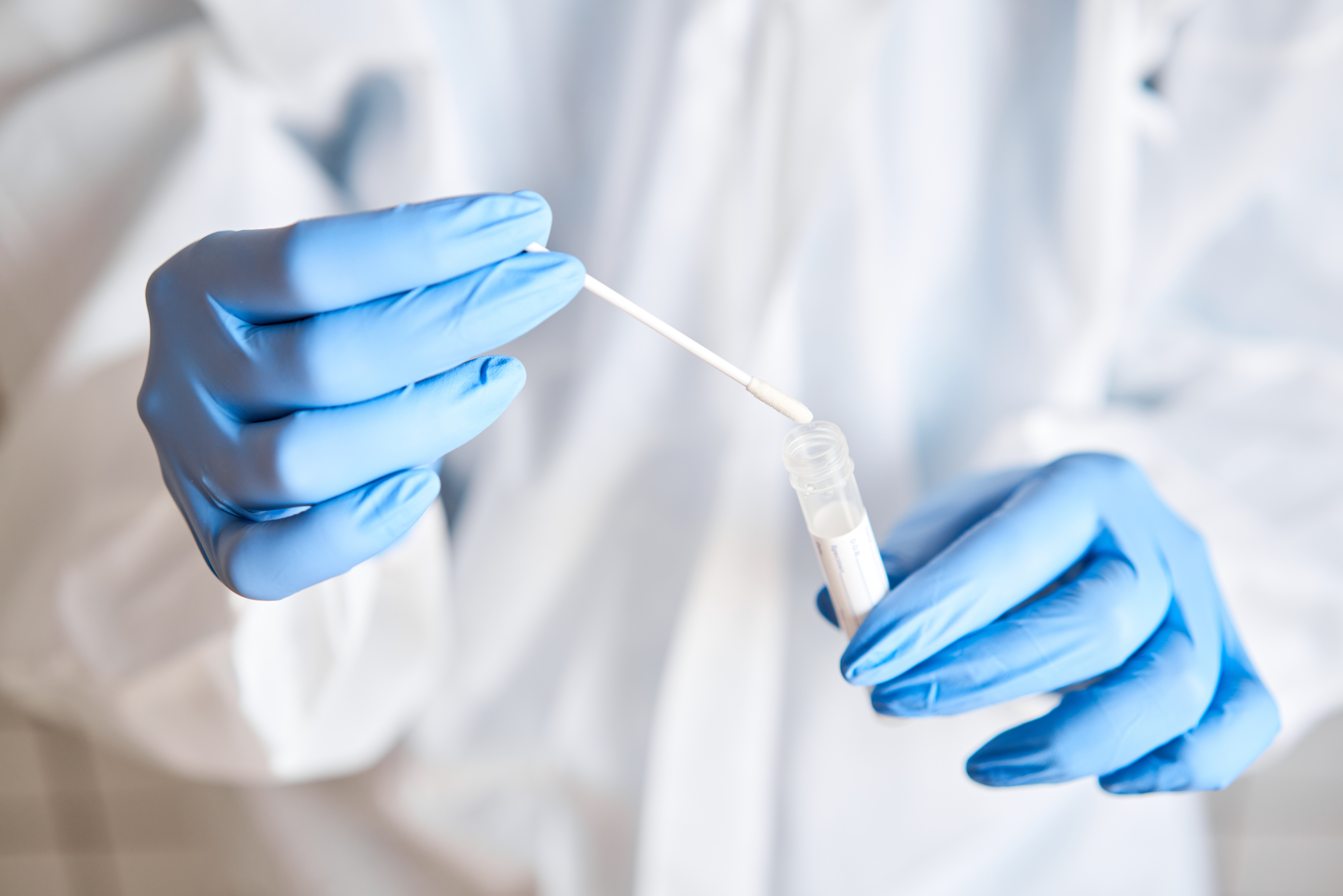
This technology enables rapid, non-invasive detection and differentiation of all four dengue virus serotypes using saliva samples, with advanced qPCR and RT-LAMP assays, making dengue diagnosis easier, faster, and more accessible without the need for blood draws. Background:
Dengue virus (DENV) is a major global health concern, with hundreds of millions of infections occurring annually, particularly in tropical and subtropical regions. Accurate and timely diagnosis is crucial for effective patient management and for controlling outbreaks, especially since infection with one DENV serotype can increase the risk of severe disease upon subsequent infection with a different serotype. Traditionally, DENV diagnostics have relied on blood-based methods, which require trained phlebotomists, specialized equipment, and can be invasive and uncomfortable for patients. This reliance on blood samples poses significant challenges in resource-limited settings, where access to healthcare infrastructure and skilled personnel may be limited, and where rapid, large-scale testing is often needed during outbreaks. Current diagnostic approaches for DENV, such as conventional PCR and serological tests, face several limitations. Blood-based PCR assays, while sensitive, often require complex sample preparation, including RNA purification, and are susceptible to inhibitors present in crude samples, which can compromise accuracy. Serological assays, on the other hand, may not reliably distinguish between DENV serotypes or between primary and secondary infections, leading to potential misdiagnosis. Furthermore, the need for cold chain storage, specialized reagents, and laboratory infrastructure restricts the deployment of these tests in field or point-of-care settings. These challenges highlight the need for more accessible, rapid, and non-invasive diagnostic solutions that can be implemented widely, particularly in outbreak-prone and resource-constrained environments.Technology Overview:
This technology provides a rapid, non-invasive diagnostic solution for detecting and differentiating all four dengue virus (DENV) serotypes using saliva samples. It integrates two advanced nucleic acid testing methods: a multiplex quantitative PCR (qPCR) assay and a reverse transcription loop-mediated isothermal amplification (RT-LAMP) assay. The multiplex qPCR can simultaneously identify DENV1–4 and a human internal control in under 90 minutes, with high sensitivity down to 3–5 viral RNA copies per microliter, and is compatible with both purified and crude saliva samples. The RT-LAMP assay, operating at a single temperature, delivers serotype-specific results in as little as 7–15 minutes and supports simple colorimetric or lateral-flow readouts, making it suitable for point-of-care settings. Both methods use standard commercial reagents and are designed for ease of use, eliminating the need for trained phlebotomists and enabling deployment in resource-limited environments. This technology is differentiated by its comprehensive approach to dengue diagnostics, leveraging large-scale genomic analysis to design highly specific primers and probes that ensure accurate serotype identification directly from saliva. Unlike traditional blood-based tests, this solution offers a non-invasive alternative that is easier to administer and more acceptable to patients, particularly in mass screening or pediatric contexts. The assays have been validated through rigorous human challenge studies and transcriptomic analyses, demonstrating comparable sensitivity and specificity to blood-based methods while also providing insights into host immune responses. Its compatibility with digital PCR and whole-genome sequencing further enhances its utility for research and epidemiological surveillance. The combination of rapid turnaround, high accuracy, non-invasive sampling, and adaptability to point-of-care use positions this technology as a significant advancement in global dengue management and public health diagnostics. https://suny.technologypublisher.com/files/sites/adobestock_349162542.jpegAdvantages:
• Non-invasive detection of all four Dengue virus serotypes using saliva samples, eliminating the need for blood draws.
• Rapid results with multiplex qPCR providing detection in under 90 minutes and RT-LAMP assays delivering results within 7–15 minutes.
• High sensitivity and specificity by targeting conserved, serotype-distinct genomic regions, with detection limits as low as ~3 copies/µL.
• Compatibility with point-of-care settings due to tolerance of crude saliva inhibitors and use of standard commercial enzymes and reagents.
• Supports multiple readout formats including fluorescent, colorimetric, and lateral-flow assays for flexible diagnostic use.
• Enables viral RNA quantification and whole-genome sequencing directly from saliva, facilitating detailed viral analysis and surveillance.
• Reduces reliance on trained medical personnel and specialized equipment, improving accessibility in resource-limited environments.
• Potential to enhance understanding of host immune responses through saliva transcriptomic analysis alongside viral detection. Applications:
• Point-of-care dengue screening
• Rapid outbreak surveillance
• At-home dengue self-testing
• Clinical trial participant monitoring
• Travel health screening Intellectual Property Summary:
Patent application filed: 63/924,381, filed on 11/24/2025
Know-how basedStage of Development:
Design of highly specific primers and probes in hand, that ensure accurate serotype identification directly from saliva. TRL level 4.Licensing Status:
This technology is available for licensing.